The Image That Shocked the Internet: Misconceptions vs. Clinical Reality
The Image That Shocked the Internet: Misconceptions vs. Clinical Reality
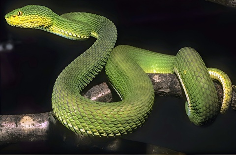
A striking image circulating on social media has captured public attention worldwide. The visual features an internal view of an organ lining that closely resembles the intricate, dark, geometric patterns of a reptile’s scales. Accompanied by sensational headlines claiming a “66-year-old’s daily habit leaves doctors shocked,” the post warns of hidden, dangerous threats lurking within our daily choices. Online discussions quickly filled with alarm, speculation, and anxiety regarding what could turn an internal organ into something resembling animal skin.
Despite the dramatic presentations found across social media platforms, this visual does not depict a mysterious modern infection, a rare mutation, or a fatal, irreversible pathology. Instead, it illustrates a classic, well-documented gastroenterological phenomenon known as Melanosis Coli.
To understand how a common lifestyle routine can transform the inner tissue of the digestive tract into a dark, patterned mosaic, it is necessary to move past clickbait headlines and examine the objective clinical facts. This comprehensive analysis will explore the physiological mechanisms, the specific compounds responsible for this structural coloration, diagnostic procedures, and how long-term digestion patterns affect human health.

What Is Melanosis Coli? Decoding the Science
Melanosis coli is a benign, non-cancerous condition characterized by dark brown or black discoloration of the mucosal lining within the large intestine. While the prefix “melan-” implies the involvement of melanin—the pigment responsible for human skin, hair, and eye color—the term is technically a misnomer. Modern histopathological investigations have established that the pigmentation is not caused by melanin at all. Instead, it is the result of a cellular byproduct called lipofuscin.
The Nature of Lipofuscin
Lipofuscin is a fine, granular, yellow-brown pigment composed of lipid-containing residues left over from lysosomal digestion. It is often referred to in cellular biology as an “aging pigment” or “wear-and-tear pigment” because it progressively accumulates in stable, non-dividing cells over time. In the case of the large intestine, however, its rapid and extensive accumulation is triggered by accelerated cellular turnover rather than natural, slow chronological aging.
Historical Context and Medical Discovery
The phenomenon was first recorded in modern medical literature in the early 19th century:
-
1830: French anatomist Gabriel Andral and pathologist Jean Cruveilhier first described a distinctive hyperpigmentation within the large bowel during autopsies.
-
1857: The prominent German physician Rudolf Virchow formally coined the term Melanosis Coli to categorize the dark, slate-gray, or brown appearance of the mucosa.
-
1928: Researcher W. E. Bartle established a definitive epidemiological correlation between this unique pigmentation and the habitual consumption of specific botanical remedies used to manage irregular digestion.
The Pathophysiology: How the Tissue Changes Color
The striking, patterned appearance observed in endoscopic photography is a direct visual representation of a cellular defense mechanism operating under metabolic stress. The transformation occurs through a precise, multi-step biological pathway within the mucosal layer of the digestive system.